
11.1. Discuss general structure of human heart (Copy)
〰️ Unit 1: General Structure of the Human Heart & The Cardiac Cycle
Chapter 11: Circulation
Student Learning Outcomes (SLO 11.1)
Learning Objectives
- Describe the anatomical location and the protective tissue layers forming the heart wall (epicardium, myocardium, endocardium).
- Identify the four chambers of the heart, the major septa separating them, and the great vessels routing blood in and out.
- Explain the structural mechanics and physiological purpose of the atrioventricular (AV) and semilunar valves.
- Provide a foundational overview of the cardiac cycle, defining systole, diastole, and the mathematical relationship of Cardiac Output.
📺 Video Lesson: Anatomy of the Heart
A comprehensive 3D medical animation depicting the intricate muscular arrangement, valve mechanics, and dual-pump blood flow of the human heart.
1. Anatomy and Protective Layers
The human heart is a highly specialized, fist-sized muscular pump located within the thoracic cavity in a central space called the mediastinum, resting on the diaphragm and flanked by the lungs. It is enclosed in a tough, double-layered sac called the Pericardium, which anchors the heart and secretes a lubricating serous fluid to prevent friction during rapid beating.
The actual wall of the heart consists of three distinct tissue layers:
- Epicardium: The thin, transparent outermost layer (also known as the visceral layer of the serous pericardium).
- Myocardium: The thick, middle layer composed entirely of cardiac muscle tissue. This layer is responsible for the powerful contractions that eject blood. The myocardium of the left ventricle is significantly thicker than the right because it must generate enough pressure to force blood through the entire systemic circulation.
- Endocardium: A smooth, innermost layer of simple squamous epithelium that lines the chambers and covers the heart valves, ensuring frictionless blood flow and preventing unwanted clot formation.

2. Chambers and the Great Vessels
The heart functions as a synchronized dual-pump system, divided into four internal chambers: two superior receiving chambers (Atria) and two inferior discharging chambers (Ventricles).
- Right Atrium: Receives deoxygenated blood returning from the systemic circulation via the Superior Vena Cava, Inferior Vena Cava, and the coronary sinus.
- Right Ventricle: Pumps deoxygenated blood out through the Pulmonary Trunk (which splits into pulmonary arteries) toward the lungs for oxygenation.
- Left Atrium: Receives freshly oxygenated blood returning from the lungs via four Pulmonary Veins.
- Left Ventricle: Pumps oxygen-rich blood forcefully out through the Aorta—the body’s largest artery—to supply the entire systemic circuit.
To prevent the mixing of oxygen-rich and oxygen-poor blood, the heart is longitudinally divided by solid muscular walls: the interatrial septum separates the atria, and the thick interventricular septum separates the ventricles.

3. The Valve System: Enforcing Unidirectional Flow
To maintain a strict one-way flow of blood and prevent fatal regurgitation (backflow), the heart relies on four pressure-sensitive mechanical valves.
Atrioventricular (AV) Valves
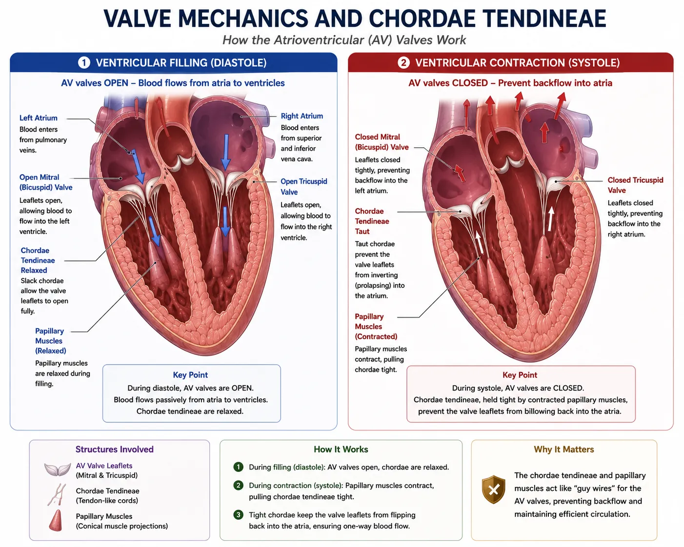
These valves lie between the atria and the ventricles. Their delicate flaps (cusps) are anchored to the ventricular walls by strong, parachute-like fibrous cords called chordae tendineae, which are attached to specialized papillary muscles. When the ventricles contract, the papillary muscles pull the cords tight, preventing the valves from prolapsing backward into the atria.
- Tricuspid Valve: Located on the right side of the heart (three cusps).
- Bicuspid (Mitral) Valve: Located on the left side of the heart (two cusps).
Semilunar Valves
These valves guard the bases of the two large arteries exiting the ventricles. They lack chordae tendineae; instead, their three half-moon-shaped pocket flaps snap shut when arterial pressure exceeds ventricular pressure.
- Pulmonary Valve: Located between the right ventricle and the pulmonary trunk.
- Aortic Valve: Located between the left ventricle and the aorta.
4. Overview of the Cardiac Cycle
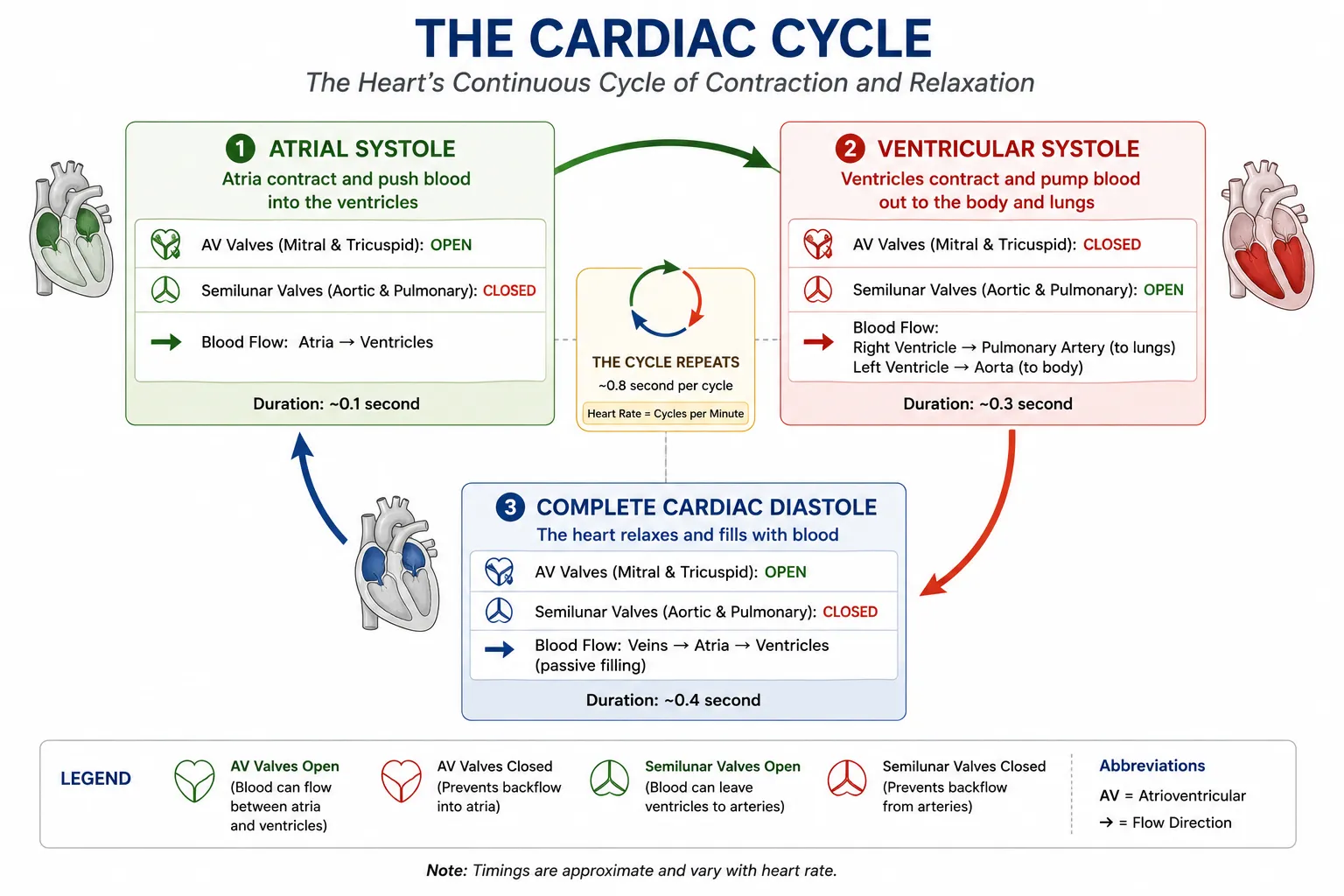
The Cardiac Cycle encompasses all the electrical and mechanical events associated with one complete heartbeat—usually lasting about 0.8 seconds in a resting adult. The cycle consists of alternating periods of contraction and relaxation:
- Systole: The active contraction phase of the heart muscle, where pressure dramatically rises to forcefully eject blood out of the chambers.
- Diastole: The passive relaxation phase, where the muscle fibers elongate, chamber pressure falls, and the heart physically refills with blood.
The efficiency of this cycle is often measured quantitatively using Cardiac Output (the total volume of blood pumped by a ventricle in one minute). Cardiac Output is mathematically defined by the equation:
$$text{CO} = text{HR} times text{SV}$$
Where $text{CO}$ is Cardiac Output, $text{HR}$ is Heart Rate (beats per minute), and $text{SV}$ is Stroke Volume (the volume of blood ejected per beat).
🎯 MDCAT Exam Insights
- Valve Location Mnemonic: Examiners often test AV valve placement. Remember the mnemonic LAB RAT: Left Atrium Bicuspid / Right Atrium Tricuspid.
- Structural Discrepancy: Be prepared to explain why the Left Ventricle has a much thicker myocardium than the Right Ventricle. The answer is purely mechanical: The right side only pumps to the adjacent lungs (low resistance), while the left side must generate massive pressure (~120 mmHg) to push blood through the entire systemic circuit (high resistance).
- Chordae Tendineae Function: Never say the chordae tendineae open the valves. They do not. They function exclusively as stabilizing guy-wires to prevent the valves from blowing backward (prolapsing) into the atria when the powerful ventricles contract.
📝 Concept Check
1. Which of the following anatomical structures is directly responsible for preventing the backflow of oxygenated blood from the left ventricle into the left atrium during ventricular systole?
The Pulmonary Semilunar Valve
The Aortic Semilunar Valve
The Bicuspid (Mitral) Valve
Check Answer
Explanation: The bicuspid valve, located between the left atrium and left ventricle, snaps shut when the left ventricle contracts, forcing blood to exit exclusively through the aortic valve.
2. The myocardium of the left ventricle is structurally much thicker and more muscular than the myocardium of the right ventricle. What is the physiological reason for this asymmetry?
The left ventricle must overcome high systemic vascular resistance to pump blood throughout the entire body.
The left ventricle pumps blood at a faster heart rate than the right ventricle.
The left ventricle is the only chamber supplied by the coronary arteries.
Check Answer
Explanation: Both ventricles pump the exact same volume of blood per beat. However, the pulmonary circuit is short and low-pressure, while the systemic circuit is vast and high-pressure. The left ventricle requires a thicker muscle mass to generate the necessary force.
3. If a patient’s resting heart rate (HR) is 70 beats per minute, and their stroke volume (SV) is 70 milliliters per beat, what is their calculated Cardiac Output (CO)?
700 mL/min
4900 mL/min
70 mL/min
Check Answer
Explanation: Cardiac output is the product of heart rate and stroke volume ($text{CO} = text{HR} times text{SV}$). Thus, $70 times 70 = 4900$ milliliters per minute (or 4.9 liters per minute), which is approximately the total blood volume of an average human!
➡ Coming Next
Unit 2: Describe the Detailed Phases of the Heartbeat
⏱️