
13.1. Discuss the functions of main part of respiratory system (Copy)
〰️ Unit 1: Functions of the Main Parts of the Respiratory System
Chapter 13: Respiration
Student Learning Outcomes (SLO 13.1)
Learning Objectives
- Differentiate between the conducting zone (anatomical dead space) and the respiratory zone.
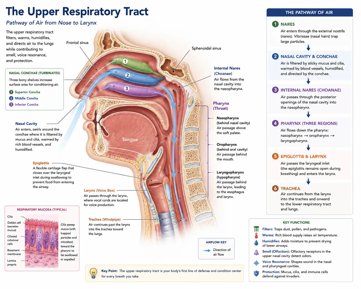
- Describe the physiological roles of the nasal cavity, pharynx, and larynx in conditioning inspired air and protecting the airway.
- Analyze the structural adaptations of the trachea and bronchial tree, specifically cartilage rings and the mucociliary escalator.
- Detail the microscopic anatomy of the alveoli, identifying the distinct functions of Type I and Type II pneumocytes.
📺 Video Lesson: Anatomy of the Respiratory System
An engaging, high-yield overview of the respiratory tract’s anatomy, demonstrating how each structural component is perfectly adapted to filter, warm, and transport air into the deep lungs.
1. The Conducting Zone (Upper Respiratory Tract)
The respiratory system is functionally divided into two zones. The Conducting Zone includes all the passageways that provide a rigid conduit for air to reach the gas exchange sites. These structures also cleanse, humidify, and warm the incoming air. Because no actual gas exchange occurs here, it is referred to as anatomical dead space (typically $approx 150text{ mL}$).
- Nasal Cavity: The primary entryway for air. It contains coarse hairs (vibrissae) to filter large particles. The nasal conchae create air turbulence, allowing a rich capillary bed to warm the air, while mucous secretions humidify it and trap fine dust and bacteria.
- Pharynx (Throat): A muscular funnel that serves as a common passageway for both food and air. It is divided into the nasopharynx, oropharynx, and laryngopharynx.
- Larynx (Voice Box): A complex cartilaginous structure. Its primary biological function is to provide an open airway and to act as a switching mechanism to route air and food into the proper channels. The epiglottis, an elastic cartilage flap, acts as a lid to cover the laryngeal inlet during swallowing, preventing food from entering the trachea.
2. The Conducting Zone (Lower Respiratory Tract)
Air passes from the larynx into the lower respiratory tract, navigating a heavily branched network of tubes often called the respiratory tree.
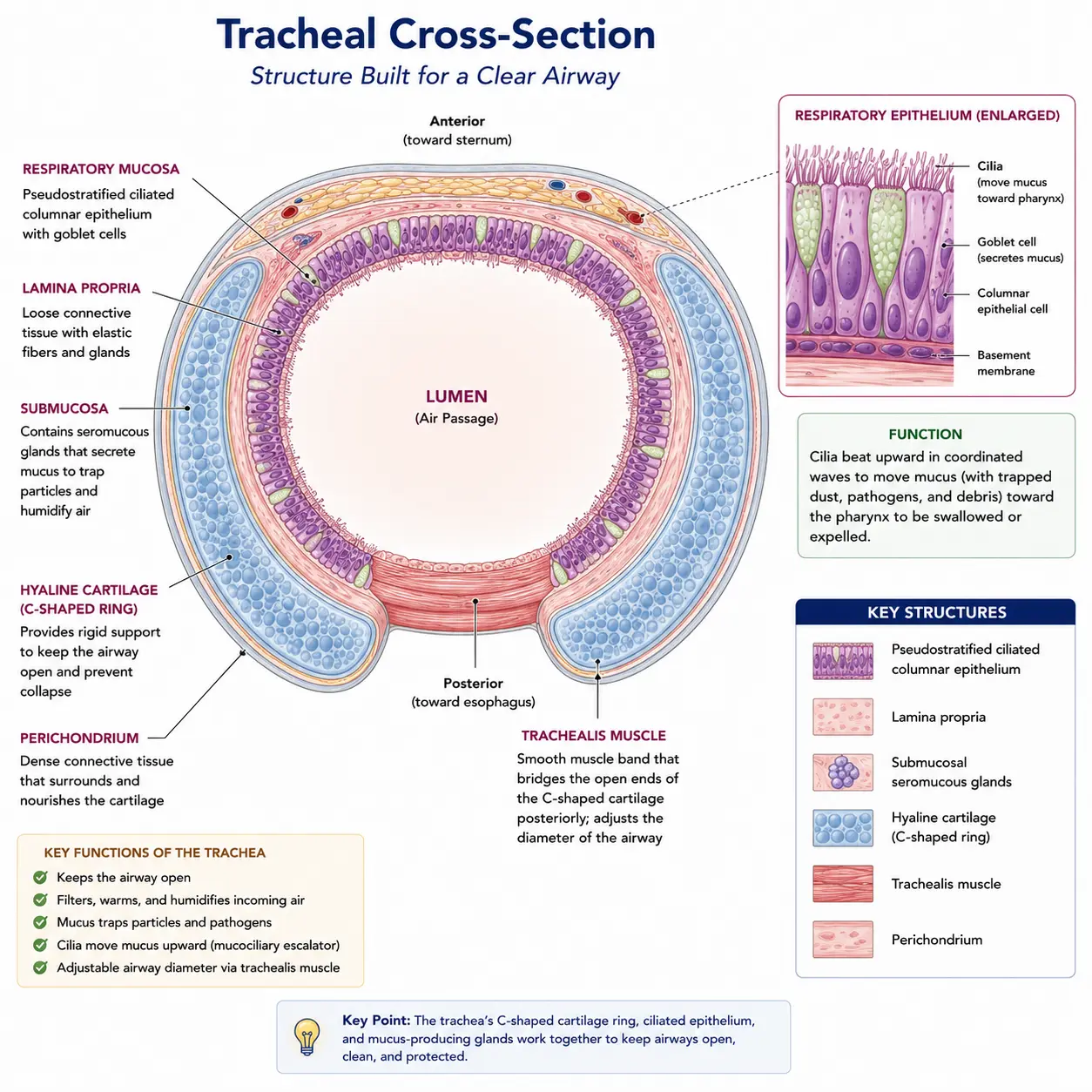
- Trachea (Windpipe): A flexible tube ($approx 10text{-}12text{ cm}$ long) that descends into the mediastinum. It is structurally supported by $16text{-}20$ C-shaped rings of hyaline cartilage. These rings prevent the trachea from collapsing under negative pressure during inhalation. The open, non-cartilaginous part of the “C” faces posteriorly, allowing the esophagus to expand into the trachea when swallowing large boluses of food.
- The Mucociliary Escalator: The inner lining of the trachea is composed of pseudostratified ciliated columnar epithelium with abundant goblet cells. Goblet cells secrete sticky mucus to trap debris, while the cilia rhythmically beat upward, moving the contaminated mucus away from the lungs toward the throat to be swallowed or spat out.
- Bronchial Tree: The trachea bifurcates into the Right and Left Primary Bronchi. These divide into secondary (lobar) bronchi, and then tertiary (segmental) bronchi. As the tubes get smaller (eventually becoming bronchioles), the cartilage rings are gradually replaced by irregular plates, and the amount of smooth muscle in the walls increases significantly, allowing for bronchoconstriction and bronchodilation.
3. The Respiratory Zone (Alveoli)
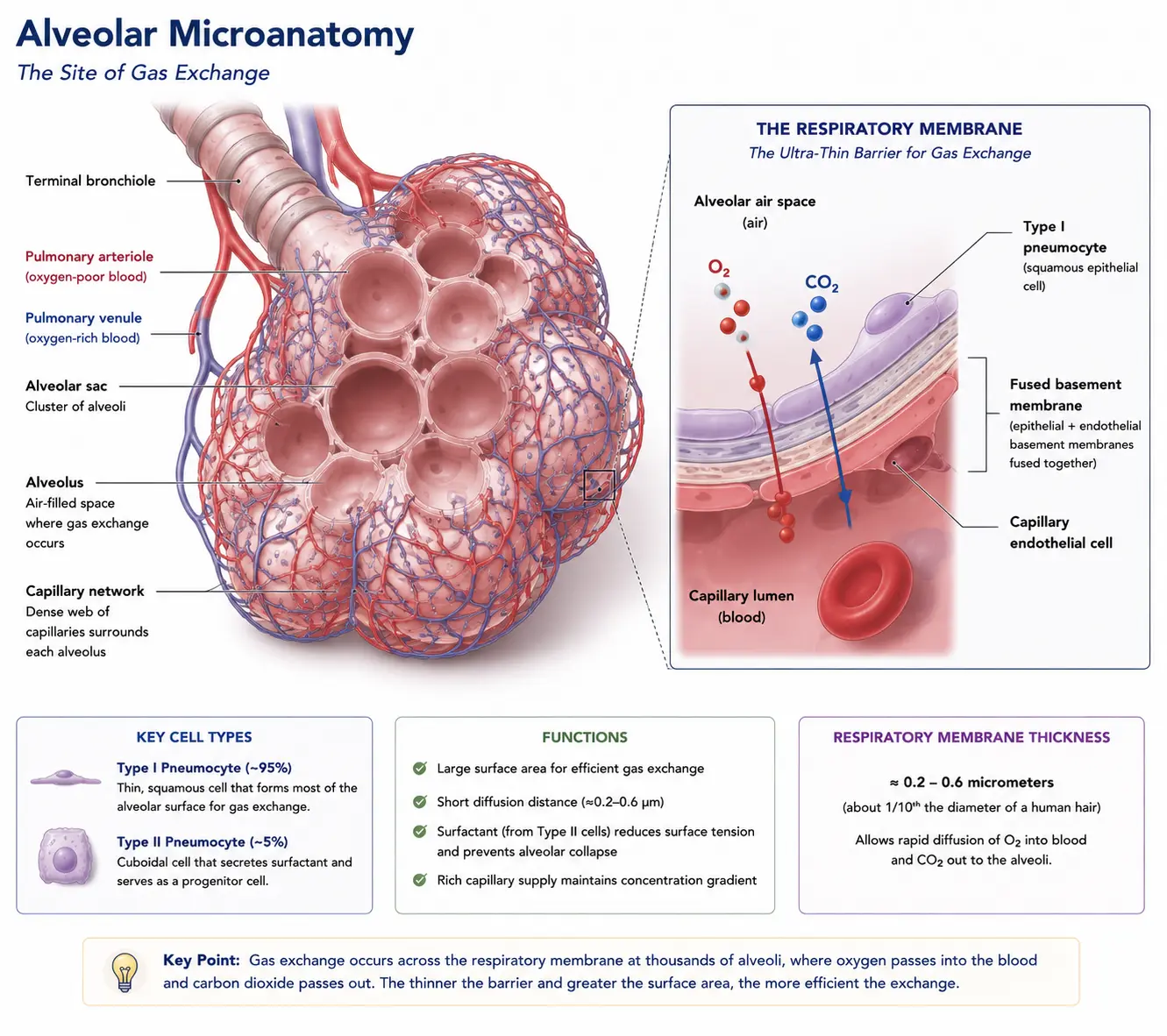
The conducting zone ends at the terminal bronchioles. The Respiratory Zone is defined by the presence of thin-walled air sacs called alveoli, which are the actual sites of gas exchange (diffusion of $O_2$ and $CO_2$).
Human lungs contain roughly 300 million alveoli, providing a massive surface area (approximately $70text{ m}^2$, or the size of a tennis court) for optimal gas diffusion. The alveolar walls are composed of two highly specific cell types:
- Type I Pneumocytes: Extremely thin, simple squamous epithelial cells that form the vast majority ($95%$) of the alveolar wall. Their thinness allows for instantaneous gas diffusion into the adjacent pulmonary capillaries.
- Type II Pneumocytes: Cuboidal cells scattered among the Type I cells. They secrete a critical detergent-like substance called surfactant (a complex of phospholipids and proteins). Surfactant coats the inner alveolar surface, drastically lowering the surface tension of water, preventing the alveoli from collapsing inward on themselves during exhalation.
4. The Lungs and Pleura
The lungs are paired, spongy organs occupying the entire thoracic cavity except for the mediastinum. Due to the presence of the heart on the left side, the lungs are asymmetrical:
- Right Lung: Larger, heavier, and divided into three lobes (superior, middle, inferior).
- Left Lung: Smaller, divided into two lobes (superior, inferior), and features a concave impression called the cardiac notch to accommodate the heart’s apex.
Each lung is enclosed in a double-layered serous membrane called the Pleura. The parietal pleura lines the thoracic wall and diaphragm, while the visceral pleura covers the external lung surface. The ultra-thin slit between them (the pleural cavity) is filled with lubricating pleural fluid, which allows the lungs to glide frictionlessly during breathing and creates the vital negative surface tension that keeps the lungs expanded.
🎯 MDCAT Exam Insights
- Cartilage Shape: Examiners often ask why the tracheal cartilage is C-shaped rather than a complete ring. The answer is always physiological: The incomplete posterior gap accommodates the expansion of the esophagus when swallowing large boluses of food, preventing the airway from acting as a rigid obstruction to digestion.
- Epithelial Transitions: You must trace the cellular changes down the respiratory tract. It begins as pseudostratified ciliated columnar in the trachea (for sweeping debris), transitions to cuboidal in the bronchioles, and finally becomes extremely thin simple squamous in the alveoli (to allow rapid gas diffusion).
- Right Primary Bronchus Trap: If a child aspirates (inhales) a foreign object like a marble, which lung is it more likely to enter? The Right Lung. The right primary bronchus is wider, shorter, and more vertically oriented than the left, making it the path of least resistance.
📝 Concept Check
1. The inner lining of the trachea acts as a highly effective “mucociliary escalator.” Which specific type of epithelial tissue forms this specialized lining?
Stratified squamous epithelium
Pseudostratified ciliated columnar epithelium
Transitional epithelium
Check Answer
Explanation: This specific tissue contains goblet cells to produce sticky mucus that traps dust and pathogens, while the apical cilia beat rhythmically upward to sweep the contaminated mucus away from the deep lungs and toward the throat.
2. What is the primary physiological function of the surfactant secreted by Type II pneumocytes in the alveoli?
To reduce the surface tension of water lining the alveoli, preventing alveolar collapse during exhalation.
To enzymatically digest inhaled airborne bacteria and viruses.
To increase the thickness of the respiratory membrane, protecting the capillaries from inhaled toxins.
Check Answer
Explanation: The thin layer of water lining the alveoli has high surface tension (due to hydrogen bonding) that constantly tries to pull the alveoli shut. Surfactant, a detergent-like lipid/protein complex, breaks these bonds, lowering the surface tension and keeping the alveoli patent (open).
3. The structural support of the human trachea relies on rings of hyaline cartilage. Why are these cartilaginous rings strictly C-shaped rather than forming complete circular rings?
To allow the anterior expansion of the spinal cord during cervical flexion.
To accommodate the expansion of the posterior esophagus when a large bolus of food is swallowed.
To provide an attachment point for the vocal cords within the larynx.
Check Answer
Explanation: The esophagus lies directly behind the trachea. If the tracheal rings were completely solid circles, food passing down the esophagus would be blocked. The open part of the “C” faces the esophagus, allowing it to bulge slightly into the tracheal space during swallowing.
➡ Coming Next
Unit 2: Discuss the Process of Gas Exchange in Human Lungs
⏱️