
14.1. Describe the parts of human digestive system (Copy)
〰️ Unit 1: Anatomy of the Human Digestive System
Chapter 14: Digestion
Student Learning Outcomes (SLO 14.1)
Learning Objectives
- Differentiate between the primary organs of the alimentary canal and the accessory digestive organs.
- Identify the four fundamental histological layers that form the wall of the gastrointestinal (GI) tract.
- Describe the gross anatomy and regional subdivisions of the stomach, small intestine, and large intestine.
- Detail the anatomical location and structural components of the liver, gallbladder, and pancreas.
📺 Video Lesson: Digestive System Anatomy
A highly detailed, visually guided tour through the entire gastrointestinal tract, highlighting the structural adaptations of each organ from the oral cavity to the rectum.
1. Organization of the Digestive System
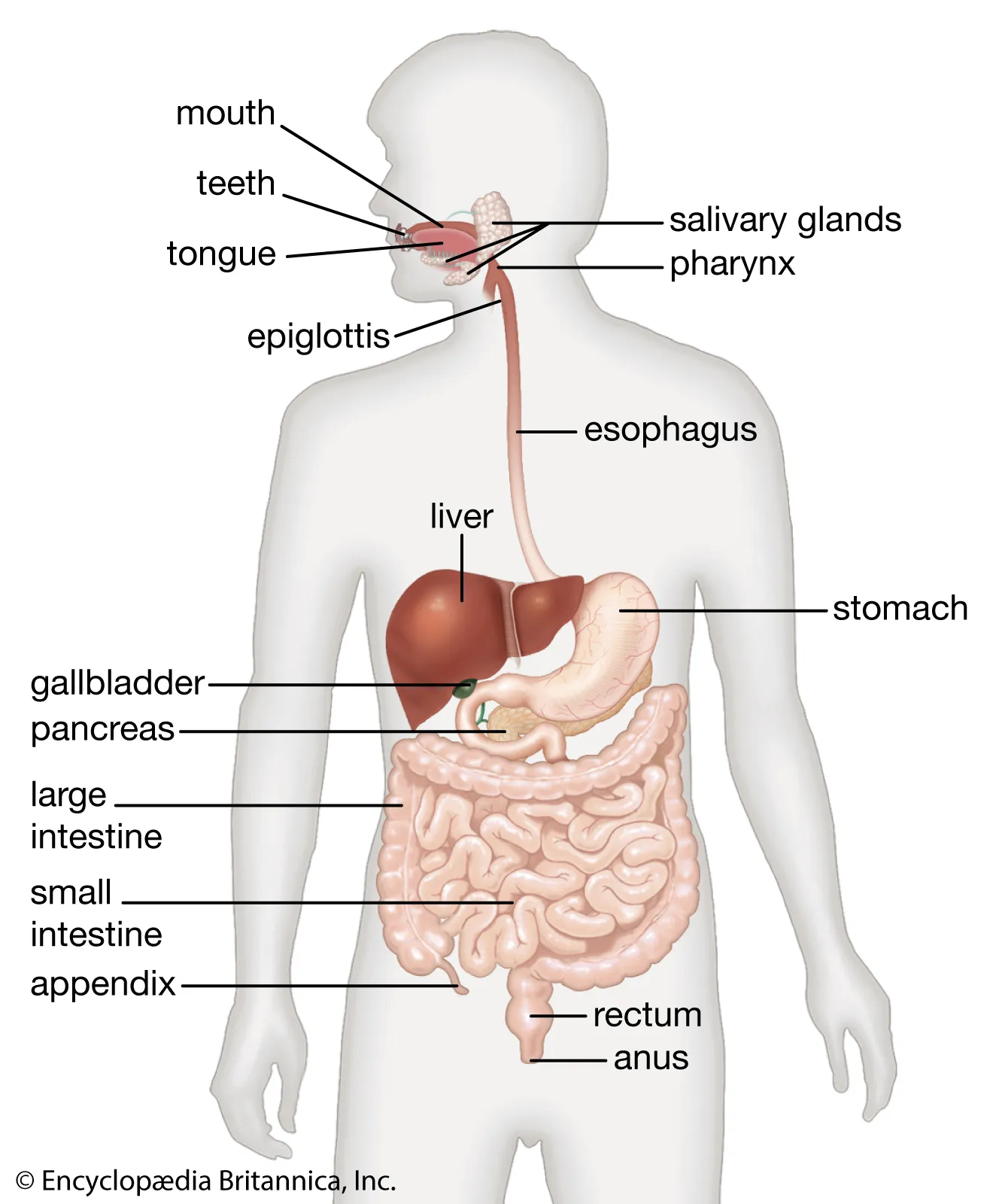
The human digestive system is anatomically divided into two main categories: the continuous tube through which food directly passes, and the supportive organs that secrete vital fluids into that tube.
- The Alimentary Canal (Gastrointestinal Tract): A continuous, coiled muscular tube measuring approximately 9 meters (30 feet) in length. It includes the mouth, pharynx, esophagus, stomach, small intestine, and large intestine.
- Accessory Digestive Organs: Structures that assist in mechanical or chemical digestion but do not form the primary tube itself. These include the teeth, tongue, salivary glands, liver, gallbladder, and pancreas.
2. Histology of the GI Tract Wall
From the esophagus down to the anal canal, the walls of the alimentary canal are remarkably consistent, consisting of four distinct, concentric tissue layers (tunics):
- Mucosa: The innermost, moist epithelial lining. It is heavily folded to increase surface area and contains glandular cells for secretion (mucus, enzymes) and absorption.
- Submucosa: A thick layer of dense connective tissue containing blood vessels, lymphatic vessels, and a major nerve network known as the submucosal (Meissner’s) plexus.
- Muscularis Externa: The smooth muscle layer responsible for segmentation and peristalsis. It typically consists of an inner circular layer and an outer longitudinal layer. It is innervated by the myenteric (Auerbach’s) plexus.
- Serosa (Visceral Peritoneum): The outermost protective layer of connective tissue and simple squamous epithelium, which anchors the tract within the abdominopelvic cavity. (Note: In the esophagus, this layer is called the adventitia).

3. Anatomy of the Upper GI Tract
The Esophagus
A straight, muscular tube approximately 25 cm long that connects the pharynx to the stomach. It pierces the diaphragm at an opening called the esophageal hiatus. Its inferior end is guarded by the lower esophageal sphincter (cardiac sphincter), which prevents highly acidic stomach contents from refluxing upward.
The Stomach
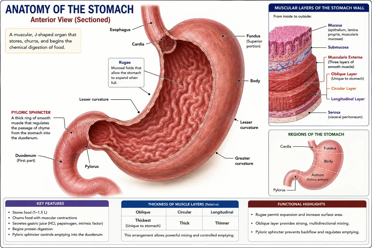
A J-shaped muscular pouch located in the upper left quadrant of the abdominal cavity. When empty, its inner mucosa collapses into large, visible folds called rugae, allowing the stomach to stretch significantly when full. Anatomically, it is divided into four regions:
- Cardia: The small region immediately surrounding the esophageal opening.
- Fundus: The dome-shaped superior portion tucked beneath the diaphragm.
- Body: The large, central midportion of the stomach.
- Pylorus: The funnel-shaped terminal end that connects to the small intestine, strictly regulated by the powerful pyloric sphincter.
4. Anatomy of the Lower GI Tract
The Small Intestine
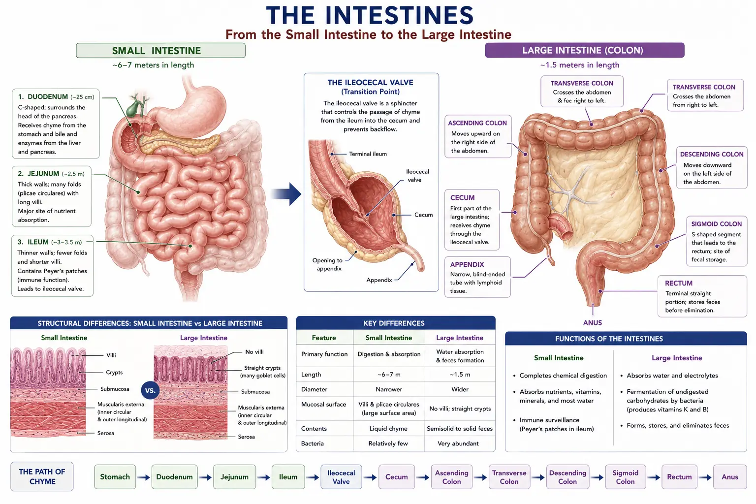
The longest segment of the alimentary canal (up to 6 meters) and the primary site of nutrient absorption. It features three subdivisions:
- Duodenum: The short (approx. 25 cm), C-shaped initial segment. It receives bile and pancreatic juice through the hepatopancreatic ampulla.
- Jejunum: The middle section (approx. 2.5 meters), responsible for the bulk of chemical digestion and absorption.
- Ileum: The final, longest section (approx. 3.5 meters), which connects to the large intestine at the ileocecal valve.
The Large Intestine
Wider in diameter but much shorter (approx. 1.5 meters) than the small intestine. Its primary role is to absorb water and package feces. It consists of:
- Cecum: A blind pouch just below the ileocecal valve, from which the vermiform appendix hangs.
- Colon: Divided into the ascending colon, transverse colon, descending colon, and the S-shaped sigmoid colon.
- Rectum & Anal Canal: The terminal holding chambers. The anal canal is controlled by an involuntary internal sphincter and a voluntary external sphincter.
5. Accessory Digestive Organs
These organs lie entirely outside the GI tract but deliver their essential secretions via specific duct systems:
- Salivary Glands: Three main pairs (Parotid, Submandibular, and Sublingual) secrete saliva to initiate carbohydrate breakdown.
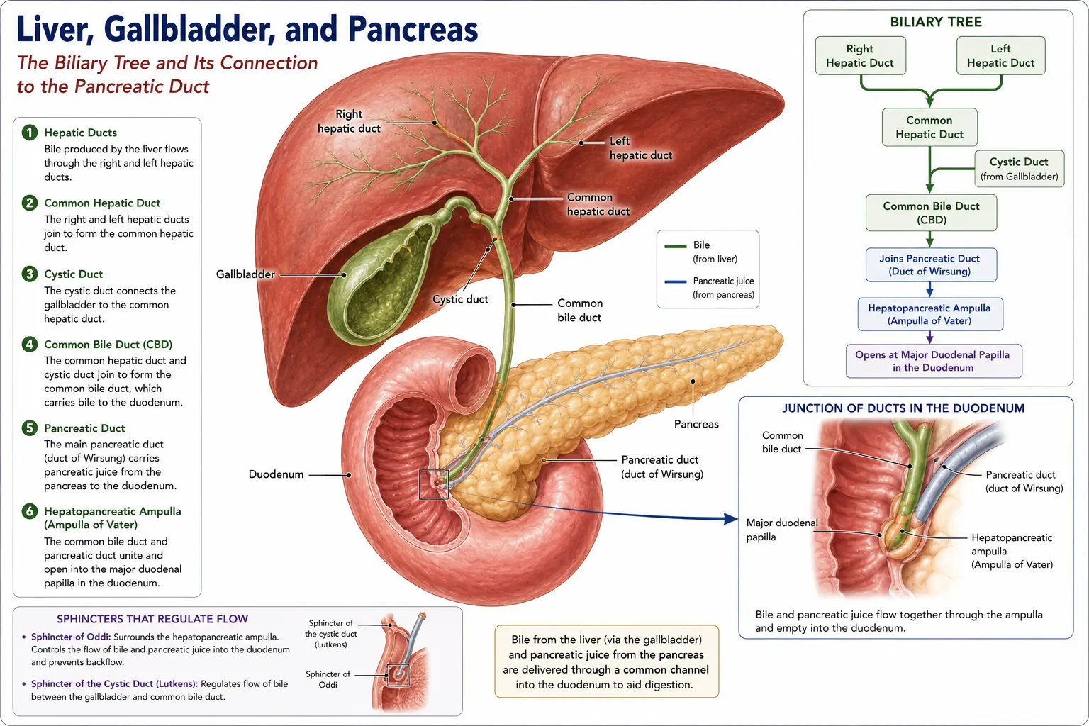
- Liver: The body’s largest internal organ, located in the upper right quadrant. It produces bile continuously.
- Gallbladder: A small, green muscular sac resting in a shallow fossa on the inferior surface of the liver. It concentrates and stores bile until it is needed for fat digestion.
- Pancreas: A soft gland lying transversely behind the stomach. Its exocrine cells produce a broad spectrum of digestive enzymes, which travel down the main pancreatic duct to join the bile duct at the duodenum.
🎯 MDCAT Exam Insights
- Histological Sequence: Exam questions frequently ask you to order the layers of the GI tract. Always remember the sequence from the lumen outward: Mucosa → Submucosa → Muscularis Externa → Serosa.
- Sphincter Identification: Entry tests often try to confuse the anatomical locations of sphincters. Remember that the Cardiac Sphincter (Lower Esophageal) is superior (near the heart), while the Pyloric Sphincter is inferior (connecting the stomach to the duodenum).
- Stomach Musculature: Most of the GI tract has two layers of smooth muscle in the muscularis externa. The stomach is the only organ that features a third, inner oblique layer, allowing it to perform aggressive mechanical churning.
📝 Concept Check
1. Which histological layer of the gastrointestinal tract is directly responsible for generating the forceful peristaltic contractions that propel food downward?
Submucosa
Muscularis Externa
Serosa
Check Answer
Explanation: The muscularis externa is composed of inner circular and outer longitudinal smooth muscle layers. Their coordinated, alternating contractions create the wave-like movements known as peristalsis.
2. Anatomically, the highly acidic contents of the stomach are prevented from violently flushing backward into the esophagus by a ring of smooth muscle known as the:
Pyloric Sphincter
Ileocecal Valve
Internal Anal Sphincter
Check Answer
Explanation: Located at the superior end of the stomach, this sphincter prevents gastric acid from refluxing into the delicate esophagus. The pyloric sphincter, on the other hand, is at the inferior end preventing rapid emptying into the duodenum.
3. The small intestine is anatomically subdivided into three distinct segments. What is the correct sequence of these segments beginning from the stomach and ending at the large intestine?
Jejunum, Duodenum, Ileum
Duodenum, Jejunum, Ileum
Ileum, Jejunum, Duodenum
Check Answer
Explanation: Food leaves the stomach and enters the short, C-shaped duodenum. It then passes into the highly coiled jejunum (the primary absorption site), and finally travels through the long ileum before entering the cecum of the large intestine.
4. Which accessory digestive organ is physically located entirely outside the alimentary canal but is directly responsible for concentrating and storing bile?
The Spleen
The Gallbladder
The Liver
Check Answer
Explanation: The liver synthesizes the bile, but it flows into the small, muscular gallbladder via the cystic duct to be stored and heavily concentrated until it is required for fat digestion in the small intestine.
5. Unlike the rest of the gastrointestinal tract, the muscularis externa of the stomach is uniquely adapted by possessing:
A thick outer layer of voluntary skeletal muscle.
Complete cartilaginous rings for structural support.
A total absence of the submucosal nerve plexus.
Check Answer
Explanation: While the entire tract relies on circular and longitudinal muscle for wave-like peristalsis, the stomach features a third, obliquely arranged layer. This allows the stomach to aggressively churn, twist, and mechanically pummel food down into a liquid paste.
6. In which histological layer of the GI tract wall would you find the Meissner’s plexus, a major nerve network controlling glandular secretions?
Submucosa
Muscularis Externa
Serosa
Check Answer
Explanation: The submucosal (Meissner’s) plexus is situated within the dense connective tissue of the submucosa, where it primarily regulates the activity of mucosal glands and local blood flow.
➡ Coming Next
Unit 2: Explain the Functions of the Main Parts of the Digestive System
⏱️