
4.1. Compare the structure of typical animal and plant cell
〰️ Unit 1: Animal vs. Plant Cell Architecture
Chapter 4: The Cell
Student Learning Outcomes (SLO 4.1)
Learning Objectives
- Identify the fundamental organelles shared by all eukaryotic cells (animal and plant).
- Detail the distinct organelles and structures exclusive to typical plant cells.
- Detail the distinct organelles and structures exclusive to typical animal cells.
- Analyze how structural differences dictate cellular functions such as cytokinesis and osmoregulation.
📺 Video Lesson: Comparing Plant and Animal Cells
A comprehensive visual comparison highlighting the structural uniqueness and shared eukaryotic machinery of both cell types.
1. Shared Eukaryotic Foundations
Both animal and plant cells are highly evolved eukaryotes. They share a massive degree of fundamental biochemical machinery required for survival. Both possess a true, membrane-bound nucleus containing linear DNA, and a suite of endomembrane system components including the Endoplasmic Reticulum (Rough and Smooth), Golgi apparatus, and various vesicles. Furthermore, both rely on mitochondria for aerobic cellular respiration (ATP synthesis) and possess a selectively permeable plasma membrane described by the Fluid Mosaic Model.
2. Exclusive Plant Cell Structures
Plant cells are generally larger ($10 \, \mu\text{m}$ to $100 \, \mu\text{m}$) and have a rigid, fixed, often rectangular shape dictated by structural adaptations for a stationary, autotrophic lifestyle:
- Cell Wall: A rigid outer layer composed primarily of cellulose microfibrils. It provides immense tensile strength, preventing osmotic lysis when the cell is in hypotonic environments.
- Plastids (Chloroplasts): Double-membrane organelles containing chlorophyll, serving as the sites of photosynthesis. Other plastids (amyloplasts, chromoplasts) store starch and pigments.
- Large Central Vacuole: Can occupy up to 90% of the cell volume. Surrounded by a specialized membrane called the tonoplast, it stores water, enzymes, and metabolic waste, while maintaining the outward turgor pressure essential for plant uprightness.
- Plasmodesmata: Cytoplasmic channels passing through the cell walls, allowing direct communication and transport between adjacent plant cells.
3. Exclusive Animal Cell Structures
Animal cells are generally smaller ($10 \, \mu\text{m}$ to $30 \, \mu\text{m}$) and lack a rigid cell wall, giving them a flexible, often irregular or highly specialized shape (e.g., neurons, erythrocytes). They possess unique features for their heterotrophic and highly mobile biology:
- Centrioles (Centrosome): Animal cells feature a pair of centrioles positioned at right angles near the nucleus. They organize the microtubule spindle apparatus essential for chromosome segregation during mitosis and meiosis. (Note: Higher plants lack centrioles but still form spindles).
- Lysosomes: Known as the “suicide bags,” these membrane-bound vesicles contain powerful hydrolytic enzymes used for intracellular digestion, autophagy, and destroying engulfed pathogens. (Rarely found in typical plant cells).
4. Critical Comparisons
The MDCAT exam requires you to distinguish between structurally similar or functionally related components:
| Comparison Pair | Key Distinctions |
|---|---|
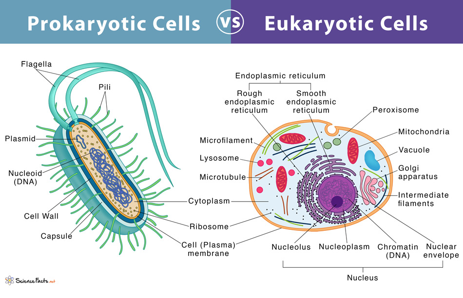
| Prokaryotic vs. Eukaryotic | Prokaryotes lack a true nucleus and membrane-bound organelles; Eukaryotes have both. |
| Peroxisome vs. Glyoxisome | Peroxisomes detoxify H2O2; Glyoxisomes (plants only) convert fatty acids to sugar. |
| Chloroplast vs. Chromoplast | Chloroplasts are green (photosynthesis); Chromoplasts are colored (fruit/flower attraction). |
| Animal vs. Plant Cell | Plants have cell walls and large vacuoles; Animals have centrioles and small vacuoles. |
🎯 MDCAT Exam Insights
- Cytokinesis Variations: Examiners frequently test how these cells divide. Animal cells perform cytokinesis via a cleavage furrow formed by a contractile ring of actin and myosin. Plant cells, unable to pinch due to the rigid cell wall, form a cell plate (phragmoplast) derived from Golgi vesicles down the cellular equator.
- The Tonoplast: Be sure to explicitly memorize the term tonoplast for the vacuolar membrane. Questions often ask which membrane is responsible for maintaining turgor pressure regulation.
📝 Concept Check
1. Which of the following organelles is prominent in animal cells and is responsible for the organization of the spindle apparatus during cell division?
Centrioles
Tonoplast
Central Vacuole
Check Answer
Explanation: Centrioles (which make up the centrosome) are unique to animal cells and lower plants, playing a crucial role in assembling microtubules for the mitotic spindle. Higher plants lack centrioles but still successfully form spindles.
2. The inability of plant cells to undergo cleavage furrow formation during cytokinesis is directly attributed to the presence of:
Chloroplasts.
A rigid cellulose cell wall.
The phragmoplast.
Check Answer
Explanation: The cell wall makes the exterior of the plant cell completely inflexible. Therefore, the cell cannot “pinch” inward (cleavage) like an animal cell. Instead, it must build a new dividing wall (cell plate) from the inside out.
3. A botanist isolates a membrane known as the tonoplast. What is the primary function of the organelle bounded by this specific membrane?
Storage of hydrolytic enzymes for apoptosis.
Generation of ATP via oxidative phosphorylation.
Maintenance of turgor pressure and storage of cellular fluids.
Check Answer
Explanation: The tonoplast is the semi-permeable membrane enclosing the large central vacuole in plant cells, which regulates ion movement and maintains the osmotic pressure (turgor) necessary to keep the plant cell firm.
➡ Coming Next
Unit 12: Prokaryotic vs. Eukaryotic Cells
⏱️