
9.1. Describe cartilage, muscle and bone
〰️ Unit 1: Description of Cartilage, Bone, and Muscle
Chapter 9: Support and Movement
Student Learning Outcomes (SLO 9.1)
Learning Objectives
- Describe the histological structure and biochemical composition of cartilage, noting its avascular nature.
- Detail the cellular components of bone tissue (osteoblasts, osteocytes, osteoclasts) and the inorganic matrix.
- Classify the three types of muscle tissue (skeletal, smooth, cardiac) based on their morphology and physiological control.
- Distinguish between compact and spongy bone structures, specifically identifying the Haversian system.
📺 Video Lesson: Tissues of the Musculoskeletal System
A microscopic and macroscopic exploration of the connective and contractile tissues that provide structural framework and movement to the human body.
1. Cartilage: The Flexible Framework
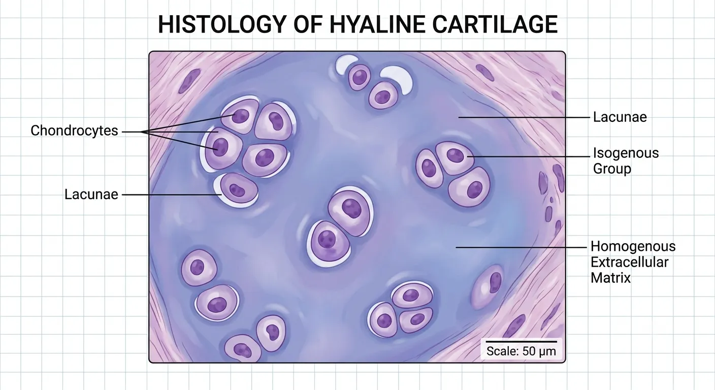
Cartilage is a specialized form of dense connective tissue that provides a smooth, resilient surface for joint movement and structural support for softer tissues (like the trachea and pinna of the ear). Its most defining physiological feature is that it is entirely avascular (lacks blood vessels) and lacks a nerve supply.
- Cells: The living cells of cartilage are called chondrocytes. They are housed in small fluid-filled cavities called lacunae.
- Matrix: Chondrocytes secrete a firm, gel-like extracellular matrix primarily composed of a protein-carbohydrate complex called chondrin, embedded with collagen and elastic fibers.
- Perichondrium: Most cartilage is surrounded by a dense layer of fibrous connective tissue called the perichondrium, from which nutrients diffuse slowly through the matrix to reach the chondrocytes.
2. Bone: The Mineralized Matrix
Bone (Osseous Tissue) is the hardest connective tissue in the body, providing rigid support, organ protection, and a mineral reservoir. Unlike cartilage, bone is highly vascularized and metabolically active.
Cellular and Chemical Composition
- Osteoblasts: Bone-forming cells that synthesize and secrete the organic portion of the matrix (osteoid).
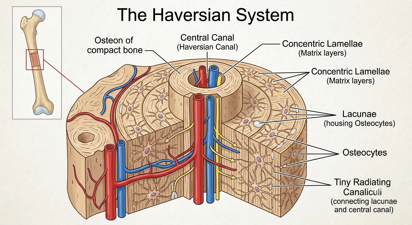
- Osteocytes: Mature bone cells derived from osteoblasts that have become trapped in their own matrix (within lacunae). They maintain daily cellular activities.
- Osteoclasts: Large, multinucleated, macrophage-like cells responsible for bone resorption (breaking down the matrix to release calcium).
- The Matrix: It is roughly $30\%$ organic (Type I collagen fibers for tensile strength) and $70\%$ inorganic. The extreme hardness comes from inorganic salts—primarily calcium phosphate, which crystallizes to form hydroxyapatite: $\text{Ca}_{10}(\text{PO}_4)_6(\text{OH})_2$.
Compact vs. Spongy Bone
Macroscopically, bone exists in two architectural forms:
- Compact Bone: The dense, solid outer layer. Its functional unit is the Osteon (Haversian System), consisting of concentric rings of matrix (lamellae) surrounding a central Haversian canal containing blood vessels and nerves.
- Spongy (Cancellous) Bone: Found at the ends (epiphyses) of long bones. It lacks true osteons and instead consists of an irregular, honeycombed network of thin bony plates called trabeculae. The spaces between trabeculae are filled with red bone marrow.
3. Muscle: The Contractile Tissue
Muscle tissue possesses the unique biological property of contractility. Muscle cells (muscle fibers) contain specialized intracellular protein filaments—primarily actin and myosin—that slide past one another to generate mechanical force. There are three distinct histological types of muscle tissue:
- Skeletal Muscle: Attached to the skeleton via tendons. It is striated (shows distinct light and dark bands under a microscope due to the highly organized sarcomeres) and is under voluntary control by the somatic nervous system. These cells are distinctly cylindrical and multinucleated.
- Smooth (Visceral) Muscle: Found in the walls of hollow organs (e.g., blood vessels, stomach, intestines). It is non-striated (smooth appearance) and is strictly involuntary, controlled by the autonomic nervous system. The cells are spindle-shaped with a single central nucleus.
- Cardiac Muscle: Found exclusively in the walls of the heart (myocardium). Like skeletal muscle, it is striated, but like smooth muscle, it is entirely involuntary. A defining feature is the presence of intercalated discs—specialized gap junctions that rapidly transmit electrical impulses, allowing the heart muscle to contract as a single coordinated unit (a functional syncytium).
🎯 MDCAT Exam Insights
- Canaliculi vs. Haversian Canals: Do not confuse these two structures in compact bone. The Haversian canal is the large central tube carrying the main blood vessels vertically. The canaliculi are the microscopic “tiny canals” radiating outward, allowing individual trapped osteocytes to pass nutrients and waste to each other via gap junctions.
- Bone Remodeling Cells: MDCAT questions frequently test the antagonistic relationship between bone cells. Remember: Osteoblasts build bone (decreasing blood calcium), while Osteoclasts consume/crush bone (increasing blood calcium under the influence of Parathyroid Hormone).
- Microscopic Muscle ID: If a histology question describes a tissue that is both “striated” and “involuntary,” it must be Cardiac Muscle. If it describes “multinucleated fibers,” it must be Skeletal Muscle.
📝 Concept Check
1. Which of the following cell types is a large, multinucleated derivative of the macrophage lineage that secretes acids and enzymes to dissolve the inorganic hydroxyapatite matrix?
Chondrocyte
Osteocyte
Osteoclast
Check Answer
Explanation: Osteoclasts are responsible for bone resorption. They secrete lysosomal enzymes to digest the organic collagen and hydrochloric acid to dissolve the inorganic calcium salts, returning $\text{Ca}^{2+}$ to the bloodstream.
2. A tissue sample viewed under a light microscope reveals elongated, spindle-shaped cells containing a single central nucleus with no visible cross-striations. From which organ was this tissue most likely extracted?
The bicep brachii attached to the humerus.
The muscular wall of the small intestine.
The compact bone of the femur.
Check Answer
Explanation: The histological description exactly matches smooth (visceral) muscle. Smooth muscle lacks the organized sarcomeres that create striations and is found in the walls of hollow autonomic organs, such as the digestive tract, blood vessels, and respiratory passages.
3. The fundamental structural and functional unit of compact bone, consisting of a central canal surrounded by concentric lamellae, is definitively known as:
An Osteon (Haversian System)
A Lacuna
A Sarcomere
Check Answer
Explanation: Compact bone is architecturally composed of millions of these cylindrical units called Osteons, which run parallel to the long axis of the bone. Spongy bone, in contrast, utilizes a network of trabeculae and lacks true osteons.
➡ Coming Next
Unit 2: Main Characteristics of Cartilage and Bone Along with Functions
⏱️